Diseases and Die-offs - More Tiger Salamanders, and an encounter with Ranavirus
A Local Tiger Salamander Die-off and Ranavirus
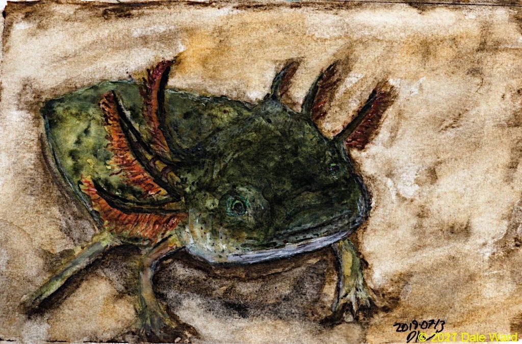 Salamander larva from the front. Pen and ink and watercolor.
Over the last month or so, I’ve been looking for more Tiger Salamander populations. One of the new Salamander ponds I’ve found is close to home, so I’ve been able to visit it repeatedly.
It’s a fascinating spot.
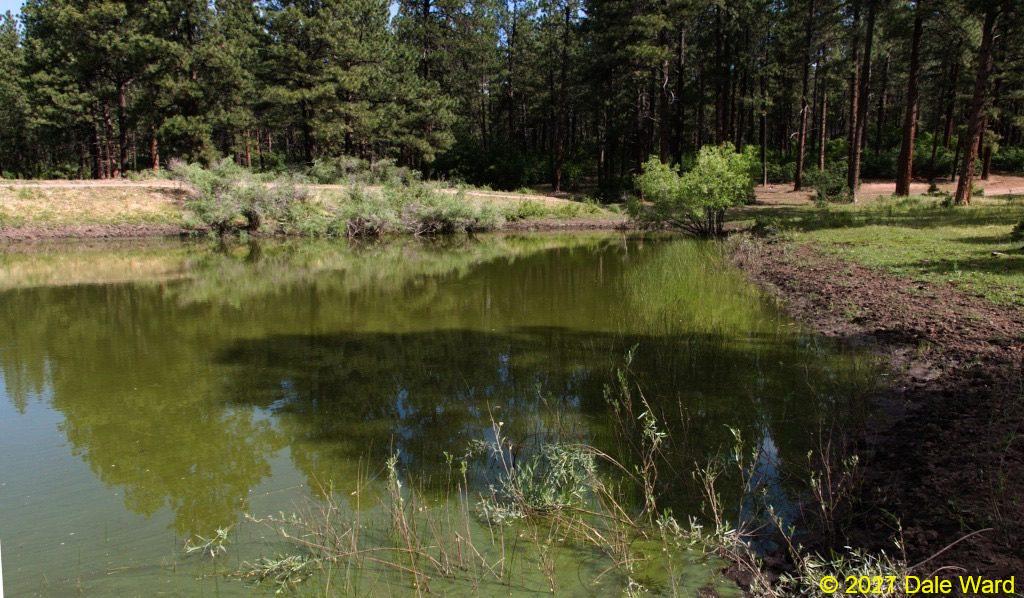 Cattle tank on the nearby San Juan National Forest. This is at the start of a big algal bloom.
For one thing, there are a lot of Tiger Salamander larvae in this pond. Walking along the edge of the pond, I’ll commonly see a larva every three or four feet. I get a little thrill with each one of them that I see - it’s like catching glimpses of tiny Crocodiles in the water. Some of them are just basking quietly in the water, others have their noses embedded in the mud as they root around and look for prey.
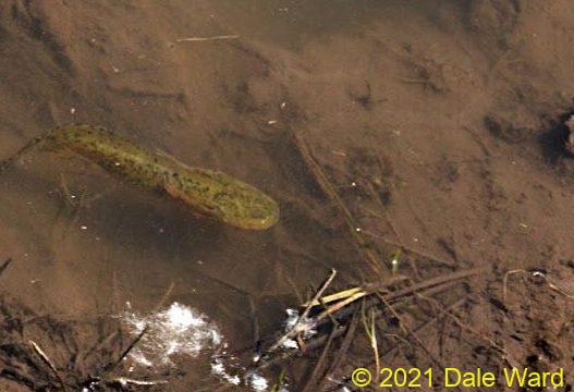
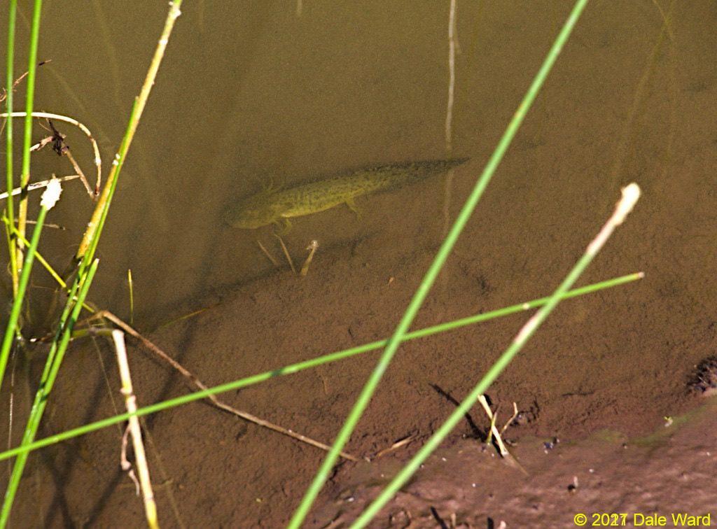 Tiger Salamander larva in the shallows of the tank. Every now and then, the larva would pulse its gills back and forth to move fresh water over its gills.
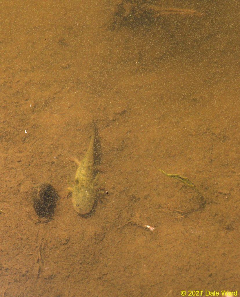 Larva basking next to a snail, in shallow waters of the stock tank.
Usually when the larvae move in the shallows, they use their legs to clamber along the bottom. This gives them a slow-motion, bouncy-looking gait, sort of like watching old movies of astronauts on the moon. When the larvae are in a hurry, though, they fold their legs and gills back against their body and swim with their tails, zipping through the water.
 Tiger Salamander larva swimming in shallow water. It’s mostly swimming by undulating its tail. Check out the way it folds its legs gills back against its body to minimize drag.
I had wanted to get a better look at the larvae and had brought a dip net with me. After a couple of false starts (and partially falling into the mud of the tank), I managed to catch a few of larvae. My first successful net-scoop, which was aimed at only one visible larva, managed to catch four of them! I hadn’t even seen the other three larvae.
Clearly, there were a lot more larvae in this tank than I thought.
I put the larvae into a small plastic aquarium that I had also brought along. I was astonished at how colorful they appeared in the clear water of the aquarium.
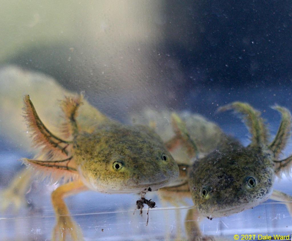 Pair of larvae in photo aquarium
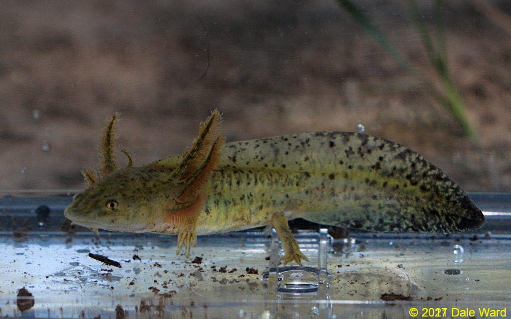
 Tiger Salamander in photo aquarium. Note the leech attached to the larva’s side. I was surprised that there were leeches around, I had assumed that the cattle tank was small and isolated enough to avoid having them.
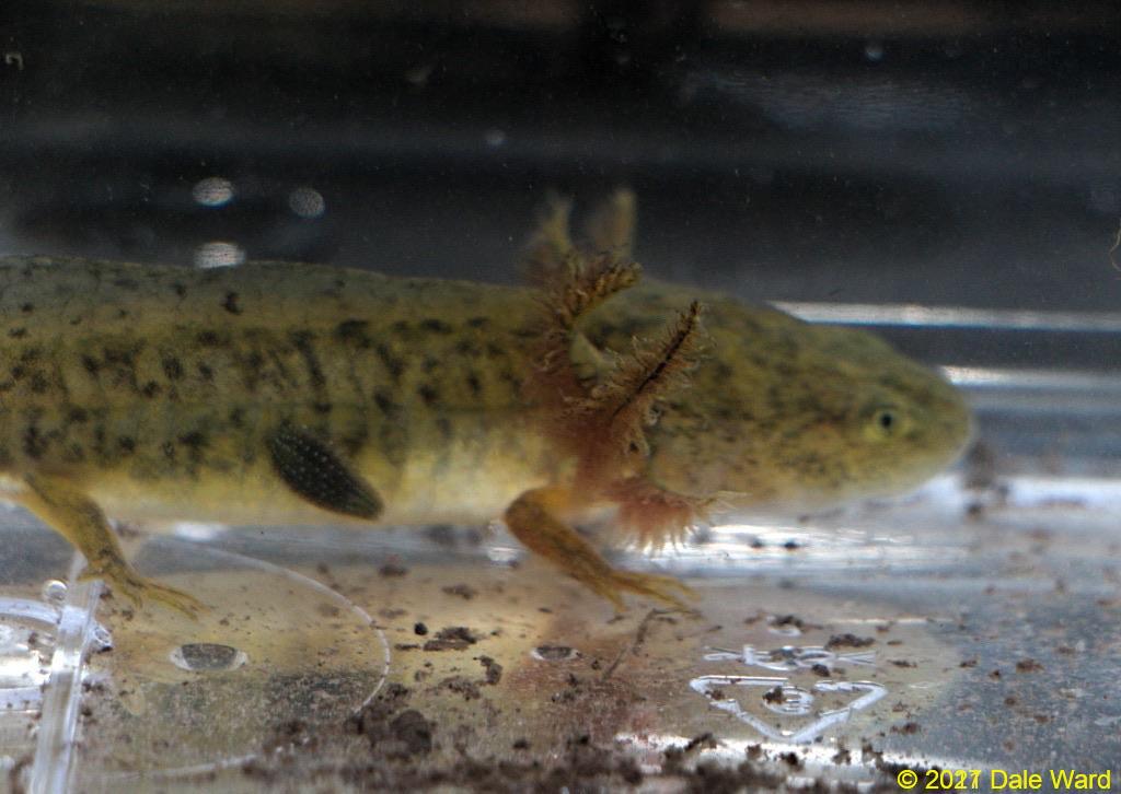
 Tiger Salamander larva in photo aquarium. The blood vessels in their gill arches are fascinating. Note especially the differences in color in the gill filaments on the foreground and the larva in the background.
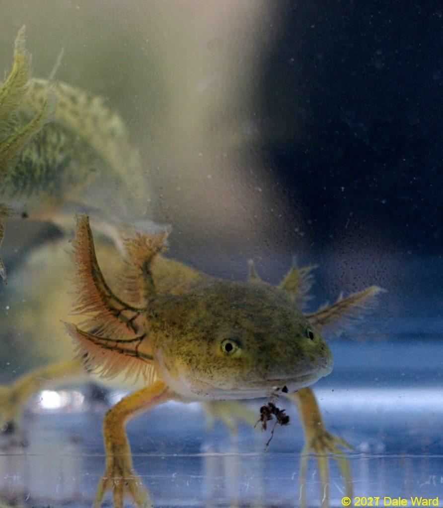
 Tiger Salamander larva in photo aquarium. Note how bright and varied the larva’s colors are in this lighting. Now it looks olive drab, with yellow and blue(!) flecks.
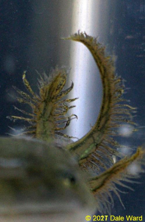 Close up of Tiger Salamander larva gills. The feathery portion is presumably where most of the gas exchange takes place. Those gill filaments change color - sometimes they are a muddy brownish color (as in this photo), and sometimes they are purplish red. Maybe they change color in response to blood oxygenation?
One of the larvae that I captured had a red, bloody-looking front leg.
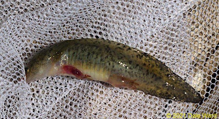 Salamander larva with injured leg.
I spoke with some Salamander friends (Daniel Spiwak and Adam Garrity) about this. They said that they have seen injuries like this caused by aggression between larvae, failed predation attempts, or even failed attempts at cannibalism.
Another interesting thing about this particular stock tank is that it is perennial - I’ve never seen it go completely dry. That makes me wonder if perhaps it has some neotenic larvae in in it as well - ones that have ‘decided’ to never metamorphose into terrestrial adults. Not sure how I’ll figure that out without some sort of trapping…
Yet another interesting thing about this stock tank was the number of dead larvae in it.
There was a period of about a week where I’d see perhaps 20 dead larvae on each of my visits. Some of the larvae would be on the bottom of the tank, others would be floating on the surface.
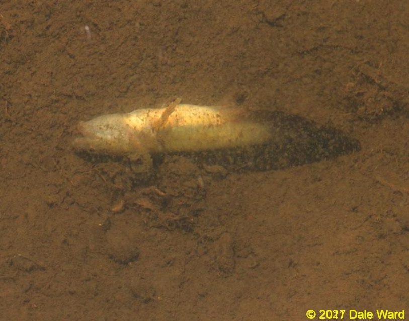 Dead Tiger Salamander larva on bottom of pool. I saw maybe 20 or so dead larvae on one of my visits.
Some of the dead larvae on the surface had rows of bloody-looking vesicles running down the sides of their bellies.
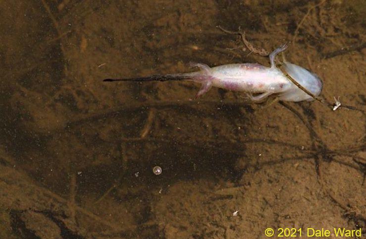 Dead Salamander larva floating on tank surface. Note the reddish vesicles on each side of the larva’s belly.
I didn’t know what could cause this, so I mentioned it to Daniel Spiwak and Adam Garrity. They suggested that it sounded like an outbreak of an Iridovirus. The Iridoviridae is family of viruses which are known pathogens of cold-blooded animals - fish, amphibians, reptiles, even insects.
Most of the ‘important’ amphibian viruses are in the genus Ranavirus. Ranaviruses are responsible for many reptile, amphibian, and fish diseases. Ranavirus are likely one of the factors causing global amphibian declines.
It turns out that there is a type of Ranavirus that hits Tiger Salamander larvae specifically - it’s named the “Ambystoma tigrinum Virus”, or “ATV” for short. From the descriptions of it that I’ve read, there’s a good chance that ATV is what was killing the larvae.
DNA work suggests that ATV originally evolved from a fish-infecting Ranavirus ancestor. The virus spreads via physical contact with infected individuals, with infected objects, or even infected water. Pond sediment can remain infectious for a relatively short while (weeks), especially if it doesn’t dry out.
Mortality from ATV can sometimes be high - I’ve read of incidents where mortality was > 90%. On the other hand, researchers have found some ponds where 60-70% of the larvae were infected with ATV, but there were no obvious mortalities.
Those low-mortality observations are in-line with what I’m seeing here - lots and lots of seemingly healthy larvae, with a few presumed victims of the virus.
This is fascinating. Many of the accounts of the virus that I’ve read have mentioned extremely high mortality rates. Note that this population hasn’t been wiped out by the virus, at least so far. There are far, far more alive and seemingly healthy larvae in the pool than there are sick ones. Why is the virus so lethal sometimes, and not others?
I wonder if the virulence of the virus has to do with how isolated the populations of larvae are. If there’s not much Salamander travel between pools, then a virus with a high virulence stands a higher chance of wiping itself out by killing too many hosts.
Paul Ewalt, in his book The Evolution of Infectious Disease, says that diseases which rely upon the mobility of their hosts for transmission would be expected to evolve so that their hosts can remain mobile.
On the other hand, diseases such as malaria, which are spread by mosquitoes, don’t rely on host mobility for transmission. In fact, immobilized hosts are easier for the vector to feed on. So we’d expect to see high virulence in a situation like that…a disease making a very, very sick host may even be preferable, since sick hosts tend not to squash mosquitoes.
Some research indicates that humans, by moving larvae from place to place, play the role of vectors with the ATV. In one of the studies that I read, the researchers tested Tiger Salamander larvae in bait shops for ATV. They found that a lot of the larvae were infected with the virus. The researchers also interviewed fishermen and bait store owners - a large proportion of them would release unused larvae to the wild.
I imagine that this would be a wonderful way to select for a highly virulent strain of virus…having humans acting as vectors. Maybe that’s why this outbreak wasn’t more lethal? Maybe it was a more ‘endemic’ type of outbreak?
 Tiger salamander with side lighting. Inktense pencils and pen-and-ink.
Complicating this question is the fact that larvae which survive an ATV infection and successfully metamorphose into adults will likely remain infectious, even though they are asymptomatic themselves.
They become carriers of the virus.
That seems to be how the virus survives from year-to-year - the breeding pools get reinfected when survivor adults return to the pool to breed. One of the studies I read found as many as 75% of the adult Salamanders returning to ponds in the Spring were infected with ATV.
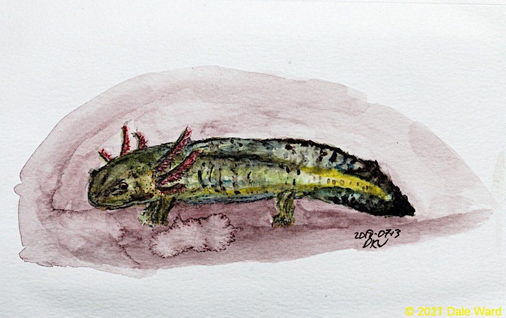
 Another attempt at capturing Salamanders with watercolors.
This is a fascinating topic, and there’s way too much of it for me to write about. Or even to make sense of, honestly.
2024 Update - Yes, this tank is positive for Ranavirus
Colorado Parks and Wildlife (CPW) is the governmental agency responsible for managing wildlife in the State of Colorado. Their Aquatic Animal Health Laboratory tested the waters in this cattle tank, the Morgan Camp Stock Tank, using eDNA (“environmental DNA”) testing. This type of test doesn’t require capturing an infected salamander. Rather, it tests whether there is DNA for the ranavirus in the water of the tank.
This tank - Morgan Camp Stock Tank, on the San Juan National Forest - was positive for ranavirus via CPW’s eDNA testing.
There can be multiple factors contributing to amphibian die-offs. This positive eDNA test does not mean that ranavirus definitely caused the die-offs that I saw. It just means that there was ranavirus DNA in the water of the tank at the time of testing.
But ranavirus was probably, at the very least, a contributing factor. And I’m betting it was one of the primary factors.
The CPW representative mentioned that there is yet another location, also near Dolores, Colorado, that tested positive for Ranavirus in 2023. CPW sequenced the DNA of that virus. It was Ambystoma Tigrinum Virus (ATV). Since that water source is relatively nearby, it’s likely that the ranavirus detected in Morgan Camp Stock Tank is also ATV.
The CPW representative said that ranavirus appears to be very widely spread in Colorado at this point, and that it’s best to assume that most water bodies in this area (Southwestern Colorado, perhaps all of Colorado) are infected with ranavirus.
And, importantly, that humans should be very careful to avoid spreading it to any potentially ranavirus-free waters.
She recommended following the Northeast Partners in Amphibian and Reptile Conservation’s protocol for disinfecting field equipment: https://northeastparc.org/docs/NEPARC-Field-Equipment-Disinfection-Protocol.pdf .
On positive note, the Morgan Camp Stock Tank tested negative for two types of chytrid fungus - Batrachochytrium dendrobatidis (Bd) and Batrachochytrium salamandrivorans (Bsal). These fungi are closely related, and have been implicated in a number of amphibian declines and extinctions.
Sources and References
Brunner, Jesse L., Danna M. Schock, Elizabeth W. Davidson and James P. Collins. 2004. INTRASPECIFIC RESERVOIRS: COMPLEX LIFE HISTORY AND THE PERSISTENCE OF A LETHAL RANAVIRUS. Ecology, February 2004.
Brunner, Jesse L, Danna M Schock and J.P. Collins. 2007. Transmission dynamics of the amphibian ranavirus Ambystoma tigrinum virus. Diseases of Aquatic Organisms 77(2), pp. 87-95.
Douglas E. Docherty, Carol U. Meteyer, Jun Wang, Jinghe Mao, Steven T. Case, and V. Gregory Chinchar. 2003. DIAGNOSTIC AND MOLECULAR EVALUATION OF THREE IRIDOVIRUS-ASSOCIATED SALAMANDER MORTALITY EVENTS. Journal of Wildlife Diseases: July 2003, Vol. 39, No. 3, pp. 556-566.
Ewald, Paul. 1996. Evolution of Infectious Disease. Oxford University Press.
Greer, Amy L., Jesse L. Brunner and James P. Collins. 2009. Spatial and temporal patters of Ambystoma tigrinum virus (ATV) prevalence in tiger salamanders Ambystoma tigrinum nebulosum. Diseases of Aquatic Organisms 85, 1-6.
Picco, A.M. and JP Collins. 2008. Amphibian commerce as a likely source of pathogen infection. Conservation Biology Dec:22(6) 1582-1589.
Price, Stephen J., Ellen Ariel, Alicia Maclaine, Gonçalo M.Rosab, Matthew J.Gray, Jesse L.Brunner, Trenton W.J.Garner. 2017. From fish to frogs and beyond: Impact and host range of emergent ranaviruses. Virology 511, November 2017. 272-279,